UnityPoint Health - John Stoddard Cancer Center - Radiation Oncology and Brachytherapy Center
Information
Number of patients waiting reflects the current number of patients waiting to be seen. This number changes frequently and is not exact.

Radiation Oncology and Brachytherapy Center
As the state's first hospital-based radiation oncology department at Iowa Methodist Medical Center, UnityPoint Health – John Stoddard Cancer Center Radiation Oncology and Brachytherapy Center continues to be one of the most technologically advanced radiation oncology departments in Iowa.
We view the family as an important part of the care we provide. Our ultimate goal is to provide accurate, precise and timely service to cure or relieve discomfort for the patient diagnosed with cancer.
Accreditations & Recognition
Stoddard Cancer Center has been recognized as an accredited breast cancer center by the National Accreditation Program for Breast Centers (NAPBC) since 2014. Receiving care at a cancer center accredited by the NAPBC means you gain access to a full range of state-of-the-art cancer care services, clinical trials and a multidisciplinary team approach to coordinate the best cancer treatment options.
Stoddard Cancer Center is an accredited cancer center by the Commission on Cancer (CoC), a program of the American College of Surgeons. CoC accreditation demonstrates a commitment to providing comprehensive, state-of-the-art cancer care close to home.
As a Clinical Radiopharmaceutical Therapy Center of Excellence, our dedicated care team boasts Radiation Oncologists certified in the interpretation of nuclear medicine studies, working hand-in-hand with certified nuclear medicine radiologists. Our team brings a wealth of experience in administering various radiopharmaceutical therapies, ensuring the highest standards of care for our patients.
Stoddard Cancer Center, one of the leading centers for radiation therapy in the Midwest, has been recognized as a Vision RT Center of Excellence. When receiving care at Stoddard Cancer Center you can expect to be treated by an oncology radiation team that has a proven expertise in the use of the AlignRT® system for all treatment sites and tattoo and mark-free radiation therapy treatment. The team demonstrates a high degree of skill and experience when delivering radiation therapy.
Stoddard Cancer Center Radiation Oncologists
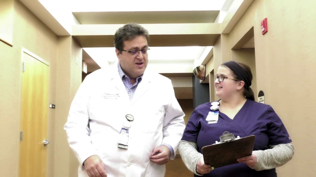
Our multidisciplinary team is available 24 hours a day, seven days a week to accommodate patients and referring physicians in the event of an emergency. Approximately 90 percent of our patients are served on an outpatient basis. Our physicians are dedicated to educating patients and their family about the treatment process every step of the way.

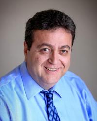
Samuel Schroeder, MD
Radiation Oncology

Anne Varner, PA
Radiation Oncology

Arshin Sheybani, MD
Radiation Oncology
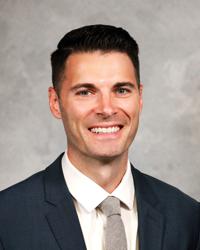
John Triantafyllos, MD
Radiation Oncology
Crosby Rock, MD
Radiation Oncology
Radiation Therapy
Radiation therapy works by focusing a beam of high-energy radiation or implanting radioactive sources in the area of tumor growth. Radiation can be used alone or in combination with other treatment, such as surgery and/or chemotherapy. In other cases, radiation therapy is employed as a supportive measure to reduce pain, pressure or bleeding. In addition, numerous benign tumors (non-cancerous) can be treated with radiation therapy.
External Beam Radiation
One of the common forms of radiation therapy used at Stoddard Cancer Center is external beam radiation therapy. This technique involves directing a "beam" of radiation from outside of your body to the cancerous organ and/or tissue within your body. External beam radiation is applied by a linear accelerator, a high-energy X-ray machine. The linear accelerator directs the radiation at the tumor. The procedure itself lasts only a few minutes. It is administered over a period of six to eight weeks, typically five days a week. Recent advances in radiation therapy can more accurately target the tumor with higher doses of radiation, while minimizing damage to healthy tissue. External beam radiation therapy poses no risk of radioactivity to you or others with whom you have contact. You can continue normal activities with family and friends.
Treatment Options
RapidArc™ radiotherapy delivers treatments using a linear accelerator. The linear accelerator rotates around the patient to deliver radiation treatments from nearly any angle. During a RapidArc treatment, radiation is shaped and reshaped as it is delivered continuously from virtually every angle in a 360-degree revolution around the patient.
Immediately prior to treatment, the exact location, size, and shape of the patient's tumor is visually observed through a simple two-minute imaging procedure using the machine's On-Board Imager. Using Image Guided Radiation Therapy (IGRT), the tumor's location is checked with a CT scan and X-ray prior to each treatment. After imaging is completed, the images are reviewed by the therapist and the patient's position can be adjusted so that an accurate treatment can be delivered. The patient does not need to move off the treatment couch for this process-all adjustments are made automatically by the treatment couch.
A RapidArc radiotherapy treatment is delivered quickly-in less than two minutes and with just one turn of the machine around the patient. RapidArc shapes and modulates a highly focused treatment beam so that it targets the tumor precisely, sparing surrounding healthy tissues. It treats the entire tumor with pinpoint accuracy and is easier on the patient, who does not have to hold still for long periods of time.
Stoddard Cancer Center had the first Intensity Modulated Radiation Therapy (IMRT) program to treat both prostate and head/neck patients in the state of Iowa. The linear accelerator delivers radiation treatments from designated angles to provide the highest level of accuracy and beam conformity by "painting" radiation doses around the tumor, lymph nodes and tissue at risk as well as shield critical structures. Similar to RapidArc, immediately prior to treatment, the exact location, size, and shape of the patient's tumor is visually observed through a simple two-minute imaging procedure using the machine's On-Board Imager. Using Image Guided Radiation Therapy (IGRT), the tumor's location is checked with a CT scan and X-ray prior to each treatment. After imaging is completed, the images are reviewed by the therapist and the patient's position can be adjusted so that an accurate treatment can be delivered. The patient does not need to move off the treatment couch for this process-all adjustments are made automatically by the treatment couch.
Stereotactic Body Radiotherapy (SBRT), initially developed in the 1990's, has been refined during recent years to treat patients with specific localized cancers who are not viable candidates for surgery. The term stereotactic refers to the ability to very precisely deliver high doses of radiation to a tumor. Because of the accuracy in delivering the radiation, the chances of killing tumor cells, while sparing healthy cells, is very high. In preparing for the treatment, detailed images of the tumor are collected.
To perform the treatment, Image Guided Radiation Therapy (IGRT) and advanced X-ray is required. Using IGRT, the tumor's location is checked with a CT scan and X-ray prior to each treatment. This technology has been in use at Stoddard Cancer Center since 2008 and in clinical trials has shown success in treating a range of localized tumors, including those in the lungs, brain, spine and pancreas.
Stoddard Cancer Center was the first to offer Stereotactic Radiosurgery (SRS) in central Iowa. SRS treats complex cancers with a precise delivery of a single, high dose of radiation in a one-day session.
Radiosurgery (one-session treatment) has such a dramatic effect in the target zone that the changes are considered "surgical." Through the use of three-dimensional computer-aided planning and the high degree of immobilization, the treatment can minimize the amount of radiation that passes through healthy tissue. Stereotactic radiosurgery is routinely used to treat brain tumors and lesions. It may be the primary treatment, used when a tumor is inaccessible by surgical means; or as a boost to other treatments for a recurring or malignant tumor. Using the TrueBeam STx, we are also able to treat cancers in the brain, lung, liver, pancreas and spine.
Stereotactic radiosurgery works the same as all other forms of radiation treatment. It does not remove the tumor or lesion, but it distorts the DNA of the tumor cells. The cells then lose their ability to reproduce and retain fluids. The tumor reduction occurs at the rate of normal growth for the specific tumor cell. In lesions such as AVMs (a tangle of blood vessels in the brain), radiosurgery causes the blood vessels to thicken and close off. The shrinking of a tumor or closing off of a vessel occurs over a period of time.
Radiosurgery versus Radiotherapy
Radiosurgery is one version of radiotherapy. Both deliver radiation but use different means and doses to minimize the risk of radiation damage to healthy tissue.
- Radiosurgery delivers radiation in single high doses that conform closely to the tumor shape, and fall off sharply at the edge of the tumor.
- Standard radiotherapy delivers small daily doses of radiation in multiple treatment sessions over several weeks.
Whether a patient receives standard radiotherapy or a form of radiosurgery depends on the type of tumor and its location.
We use AlignRT®, the premier radiation therapy guidance system, that has been shown to reduce radiation exposure to your heart. When treating cancer with radiation therapy, our goal is to deliver radiation to the tumor while protecting surrounding healthy tissue from exposure and potential damage. We take extra precautions to make sure that your heart receives minimal radiation exposure during your treatment. With AlignRT, we're using the latest technology to protect your heart while treating your cancer. No tattoo is required with AlignRT because an infrared camera outlines the surface.
In certain locations in the body, such as the lungs, tumors can move as the patient breathes. In the past, this movement has hindered doctors ability to precisely map the tumor location and to accurately deliver radiation therapy. By integrating respiratory gating software into the treatment plan, doctors can define a physical window similar to a baseball strike zone, and quickly take tumor images or deliver radiation only when the tumor passes through that region.
Respiratory gating is often combined with various treatment delivery techniques as mentioned above to deliver precise doses of radiation to tumor sites. This is particularly useful for tumors near the lungs, where radiation-induced scarring can impair future breathing.
Deep Inspiration Breath Hold (DIBH) entails taking a deep breath and holding it during treatment, allowing a decreased dose of radiation to the heart and lungs. Your radiation oncologist will review your plan to determine if this is right for you.
High Dose Rate (HDR) Brachytherapy
High Dose Rate (HDR) Brachytherapy is a form of radiotherapy where a radioactive source is brought to a tumor target via a catheter that is placed within a natural or created channel. HDR treatments are typically outpatient procedures that are much more efficient and effective than traditional therapies. The ease of catheter placement and fast delivery of the dose makes this a viable treatment option.
A typical HDR treatment lasts from 8 to 20 minutes. Stoddard Cancer Center has one of the state's only high dose rate HDR units allowing these types of treatments to be given on an outpatient basis. Patients experience fewer side effects because less radiation reaches surrounding healthy organs and tissue. Treatment is tailored to each patient based on treatment site.
Benefits
- Reduces treatment time to days instead of weeks.
- Provides fewer side effects by placing radiation directly into the tumor.
- Offers greater control and accuracy of treatment, including dosing, source of radiation location and time it stays at each location.
- Requires minimal recovery time, allowing you to go home within hours after treatment with few restrictions.
- Preserves internal tissues, as no radioactive seeds migrate into other organs.
- Able to shape radiation dose to fit the tumor.
- Allows completion of radiation before chemotherapy begins, if required.
HDR has been a proven standard of care for breast (APBI), cervical, endometrial, esophageal, prostate, rectal and lung cancer treatments. Having one of only a few HDR units in the state of Iowa for the past ten years, Stoddard Cancer Center has cared for hundreds of patients who would have otherwise had weeks of additional radiation treatment or required a hospital stay.
Radiopharmaceutical Therapy
Radiopharmaceutical therapy is a type of treatment that involves the use of radioactive substances to damage or destroy cancer cells. Upon entering the body, the radiopharmaceutical travels through the bloodstream and binds to cancer cells. This in turn delivers targeted radiation directly to the cancer cells, minimizing damage to surrounding healthy tissues.
Hyperthermia Therapy
Hyperthermia is a therapy used to heat tumors. Research has shown that in some tumors, heat can damage cancer cells and increase the effect of radiation therapy. Using focused microwave energy, the tumor is heated to approximately 108°F. Performed superficially or via interstitial probes, heat can damage cancer cells at levels that are usually safe for normal cells, and can be used to attack cancer in four major ways:
- Heat damages or weakens the cells of the tumor.
- Heat increases blood flow through the weakened tumor, which can allow radiation therapies to permeate the tumor, not just attack it from the outside.
- Increased blood flow raises oxygen levels in tumors so that the cancer can be more effectively treated by radiation therapy.
- When the body senses fever it can stimulate the natural immune system. For these reasons, hyperthermia is usually used in combination with radiation therapy.
Hyperthermia treatments are typically given several times a week, either before or after radiation therapy. Each treatment session lasts for approximately one hour. This type of treatment has shown to be most effective in treating some tumors that are recurrent or progressive despite conventional therapy. Hyperthermia can potentially be used to treat cancers which invade the skin or cancers which lie close to the surface of the skin. These can include recurrent chest wall cancer, recurrent head and neck cancer, recurrent melanoma and recurrent sarcoma.
Optune
Optune is a wearable, portable, FDA-approved glioblastoma (GBM) treatment for adult patients aged 22 years or older. It works by creating Tumor Treating Fields (TTFields), which are electric fields that slow down or stop GBM cancer cell division.
SpaceOAR Hydrogel
SpaceOAR Hydrogel is a polyethylene glycol (PEG) based hydrogel clinically shown to minimize urinary, sexual and bowel side effects and protect quality of life for prostate cancer patients undergoing radiation therapy. It provides space between the rectum and the prostate, reducing high dose radiation to the rectum.
PSMA PET Imaging
The PSMA PET scan is a test that can help your doctor learn if and where prostate cancer has spread outside your prostate gland, including to your lymph nodes, other organs, or bones. PET scans are a type of imaging test that use special dye with radioactive tracers to make cancer cells show up more clearly.
Cerianna PET Imaging
Cerianna PET scan is a test that can help your doctor learn if and where breast cancer has spread, including to your lymph nodes, other organs, or bones. This scan enables a non-invasive, comprehensive assessment of the whole-body in an individual with ER+ lesion status.
Services Offered
-
Cancer Care
Find the highest quality care from our experienced team of oncology and cancer specialists at UnityPoint Health.Cancer Care